How Fluoride Protects and Repairs Your Enamel
July 27, 2026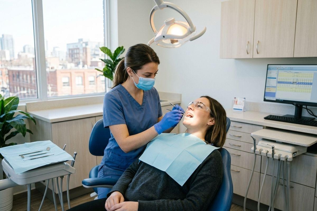
How Cavities Form and Progress
Key Takeaways
- Plaque bacteria convert sugars into acid, which lowers oral pH and weakens enamel over time.
- Demineralization is the process by which acid dissolves minerals from enamel, and it can begin before any visible hole forms.
- Early white spot lesions can be arrested with fluoride and improved hygiene, but that window closes once decay breaks through enamel.
- Cavities progress through four stages, each requiring a different level of care, from fluoride therapy to root canal treatment.
- Routine exams and x-rays allow the dental team to catch decay before symptoms appear.
What Actually Happens When a Cavity Forms
A tooth looks solid, but the process that leads to a cavity begins at the microscopic level, long before any ache or sensitivity appears. Most people associate cavities with sugar, and that connection is accurate, though the mechanism behind it is worth understanding clearly. Liberty Village Dental provides comprehensive care at every stage of this process, from early detection through restorative treatment.
For patients who want a closer look at what that care involves, the general dentistry services available at the clinic address everything from routine exams to cavity management.
Cavities form when bacteria in dental plaque metabolize sugars and release acid, which gradually dissolves the mineral structure of tooth enamel through a process called demineralization.
The Role of Plaque Bacteria & Sugar
Dental plaque is a soft, sticky film that continuously forms on tooth surfaces. It harbours a community of bacteria that feed on fermentable carbohydrates, primarily sugars. When those bacteria metabolize sugar, they release lactic acid as a byproduct.
That acid lowers the oral pH below 5.5, which is the threshold at which enamel begins to lose its mineral content. A single sugary meal or drink creates an acid exposure window of roughly 20 to 40 minutes. Frequent snacking or sipping throughout the day means the pH rarely has a chance to recover, and the enamel takes repeated hits.
Demineralization Explained
Demineralization refers to the dissolution of calcium and phosphate ions from the enamel crystalline structure caused by acid contact. The enamel surface may still look intact, but beneath it, the mineral density is declining.
The first visible sign is a chalky white spot on the enamel surface. There is no cavity yet in the conventional sense, no hole, no breakdown of the surface layer, but the structural integrity of that area has already been compromised. This is the earliest detectable stage of the caries, or tooth decay, process.
The Demineralization & Remineralization Balance
How the Mouth Naturally Repairs Itself
The mouth has a built-in repair mechanism. Saliva is slightly alkaline and carries calcium and phosphate ions that can redeposit into softened enamel, a process called remineralization. When oral pH rises back above 5.5 between meals, remineralization has the opportunity to partially counteract earlier acid damage.
Fluoride accelerates this process. It incorporates into the enamel crystal lattice and forms fluorapatite, a compound that is more resistant to acid than the original hydroxyapatite mineral structure. This is why fluoride toothpaste and professional fluoride treatments play a direct clinical role in caries prevention, not just general oral hygiene.
Why White Spot Lesions Can Still Be Arrested
At the white spot stage, the enamel surface layer remains physically intact. The subsurface has lost mineral density, but the outer shell has not collapsed. This means fluoride ions can still penetrate and support remineralization of the affected zone.
With consistent fluoride application and a reduction in acid exposure frequency, these early lesions can be arrested and, in some cases, partially recovered. Once acid erosion breaks through the enamel surface, however, the structural integrity is lost and remineralization alone cannot close that gap. At that point, restorative treatment becomes necessary.
The 4 Stages of Cavity Progression
Tooth decay does not jump from healthy enamel to a painful emergency overnight. It moves through distinct stages, and the appropriate treatment depends entirely on how far the process has advanced.
- Stage 1 involves acid erosion within the enamel layer, which may be visible as a white spot or detected by x-ray before the surface breaks down.
- Stage 2 occurs when decay reaches dentin, the softer, more porous tissue that lies beneath the enamel. Dentin contains microscopic channels called dentinal tubules that transmit sensation toward the nerve, which is why temperature sensitivity often begins at this stage.
- Stage 3 develops when decay advances toward the pulp, the inner chamber of the tooth that contains blood vessels and nerve tissue. Pulp involvement typically produces persistent discomfort and heightened sensitivity to hot and cold.
- Stage 4 represents pulp infection or abscess, where bacterial presence within the pulp has caused significant tissue damage and requires immediate attention to preserve the tooth.
When Each Stage Needs Treatment

Early Stage: Fluoride & Hygiene
White spot lesions confined to the enamel layer respond well to fluoride therapy. Professional fluoride varnish applied at a dental visit delivers a concentrated dose directly to the affected area. Combined with fluoride toothpaste used twice daily at home, this approach can halt further mineral loss.
Diet plays a direct role here. Reducing the frequency of sugar and acidic food or drink intake limits how often oral pH drops below the critical threshold. Spacing meals and avoiding constant snacking gives saliva time to neutralize acid and support natural remineralization between exposures. Oral health guidelines for adults consistently identify diet frequency as one of the most controllable factors in caries risk.
Mid Stage: Restorative Treatment
Once decay has penetrated the enamel and reached the dentin layer, fluoride therapy alone is no longer sufficient. The structural loss requires physical restoration. A dentist removes the decayed tissue and places a filling material to seal the area and protect the remaining tooth structure. Tooth-coloured composite fillings are a common option at this stage, blending with the natural tooth while providing durable coverage.
Addressing dentin-level decay promptly matters because dentin is significantly softer than enamel and decay progresses through it faster. A filling placed at this stage can protect the pulp from future involvement.
Late Stage: Pulp Involvement & Root Canal Therapy
When bacteria reach the pulp, the infection within that nerve-rich chamber produces pronounced discomfort, sensitivity that lingers after the stimulus is removed, and sometimes visible swelling. Root canal therapy, also called endodontic treatment, involves removing the infected pulp tissue, cleaning and shaping the internal canals, and sealing the tooth to prevent reinfection.
This is not a last resort. It is a clinically appropriate treatment that can preserve the natural tooth structure and prevent the infection from spreading to surrounding tissue. Prompt care at this stage avoids extraction in many cases.
Prevention & Early Detection at a Dental Practice
Routine Exams & X-Rays
One of the most consistent advantages of regular dental visits is access to diagnostic tools that identify decay before it causes symptoms. Bitewing x-rays, a standard type of dental radiograph, reveal decay developing between teeth and beneath existing restorations where a visual exam cannot reach.
The dental team can detect early-stage lesions during a routine check-up and recommend the least invasive intervention available at that point. Catching decay at Stage 1 or Stage 2 rather than Stage 3 or 4 significantly reduces both the complexity of treatment and the time involved.
Preventive Care Between Visits
Daily habits carry the majority of the preventive load between appointments. Fluoride toothpaste used twice daily, consistent flossing to remove plaque from proximal surfaces where the toothbrush cannot reach, and a diet with limited frequency of sugary or acidic foods all reduce the conditions that allow the caries process to take hold.
A dentist in Liberty Village can help tailor a preventive plan based on individual risk factors, including saliva composition, diet patterns, and existing tooth anatomy. Prevention is not one-size-fits-all, and a plan built around a person’s specific oral environment tends to be more effective than general guidelines alone.
Liberty Village Dental supports patients through every stage of this process, from early detection and fluoride therapy to restorative and endodontic care. Scheduling a routine exam is a straightforward step toward catching any developing issues while treatment options remain simple. Contact our team to arrange an appointment.